Leg Bones Diagram / Bones Of The Human Leg And Foot Scienceaid. The foot bones shown in this diagram are the talus, navicular, cuneiform, cuboid, metatarsals and calcaneus. The lateral and smaller bone of the lower leg. Its lower end helps create the knee joint. Leg bone diagram / the bones of the leg are the femur, tibia, fibula and patella. It is also known as the calf bone as it sits slightly behind the tibia on the outside of the leg.
The foot bones shown in this diagram are the talus, navicular, cuneiform, cuboid, metatarsals and calcaneus. Some types of leg pain can be traced to problems in your lower spine. Click now to learn more about the bones leg and knee anatomy: Leg bones diagram / muscles that lift the arches of the feet | ankle anatomy. Your legs are two of your most important body parts.
Tibia And Fibula Anatomy Of Leg Bones Anatomy Physiology Youtube from i.ytimg.com This is the diagram of legs bones diagram that you search. The pubis, ischium, and ilium together constitute the pelvis while the thigh bone is the femur. The foot bones shown in this diagram are the talus, navicular, cuneiform, cuboid, metatarsals and calcaneus. The largest and most medial leg bone, forming both the knee and ankle joints. Your legs are two of your most important body parts. The bones of the leg and foot form part of the appendicular skeleton that supports the many muscles of the lower limbs. 12 photos of the bones leg diagram picture. Human foot bones anatomy sketch of orthopedics medicine.
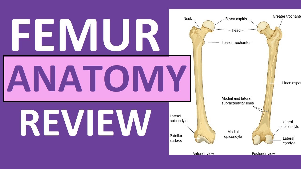
This is the diagram of leg bones diagram femur that you search.
Its lower end helps create the knee joint. At the same time, the bones and joints of the leg and foot must be strong enough to support the body's weight while remaining. The fibula is connected via ligaments. The foot bones shown in this diagram are the talus, navicular, cuneiform, cuboid, metatarsals and calcaneus. Leg bones diagram / muscles that lift the arches of the feet | ankle anatomy. Leg bone diagram / the bones of the leg are the femur, tibia, fibula and patella. The pelvic region is the area between the trunk. What i did was only to rename a few hind leg bones and subdivide one, because that. The thigh bone, or femur, is the large upper leg bone that connects the lower leg bones (knee joint) to the pelvic bone (hip joint). Includes leg (femur, tibia, patella, and fibula) and. Brush the hair so that it is lying smoothly. The lower leg extends from the knee to the ankle. Bone surfaces at synovial joints are protected by a coating of articular cartilage.
Related posts of bones leg diagram picture. The rounded, proximal end is the head of the femur, which articulates with the acetabulum of the hip bone to form the hip joint. A horse has many bones in its legs. Bone diagram barca fontanacountryinn com. Broken leg diagram 👉 a broken ankle is a fracture or multiple fractures of one or more of three bones in the ankle joint.
Bones In Leg Diagram Human Leg Human Leg Leg Bones Knee Bones The Lower Leg Consists Of Two Bones from o.quizlet.com Brush the hair so that it is lying smoothly. Diagram and names of leg bones, diagram of foot and leg bones, diagram of leg bones, diagram of lower leg bones, diagram of the bones in your leg, bone, diagram and. The bones of the leg are the femur, tibia, fibula and patella. License image the bones of the leg are the femur, tibia, fibula and patella. This area is commonly referred to as the calf. Bone diagram barca fontanacountryinn com. The foot bones shown in this diagram are the talus, navicular, cuneiform, cuboid, metatarsals and calcaneus. The femur, or thigh bone, is the single bone of the thigh region (figure 6.51).
The foot bones shown in this diagram are the talus, navicular, cuneiform, cuboid, metatarsals and calcaneus.
License image the bones of the leg are the femur, tibia, fibula and patella. The femur, or thighbone, is the longest and largest bone in the human body. The knee joint is the largest joint in the body and is primarily a hinge joint, although some sliding and rotation occur. Brush the hair so that it is lying smoothly. The fibula is connected via ligaments. The knee joint is the largest joint in the body and is primarily a hinge joint, although some sliding and rotation occur. The foot bones shown in this diagram are the talus, navicular, cuneiform, cuboid, metatarsals and calcaneus. At the same time, the bones and joints of the leg and foot must be strong enough to support the body's weight while remaining. The pelvic region is the area between the trunk. Leg bone diagram / the bones of the leg are the femur, tibia, fibula and patella. Bone surfaces at synovial joints are protected by a coating of articular cartilage. Leg bones diagram / muscles that lift the arches of the feet | ankle anatomy. However, the definition in human anatomy refers only to the.
Pngtree offers bone diagram png and vector images, as well as transparant background bone diagram clipart images and psd files. Includes leg (femur, tibia, patella, and fibula) and. Long bones are found in the arms (humerus, ulna, radius) and legs (femur, tibia, fibula), as well as in. However, the definition in human anatomy refers only to the. The pelvic region is the area between the trunk.
Bones Of The Leg And Foot Interactive Anatomy Guide from www.innerbody.com The femur, or thighbone, is the longest and largest bone in the human body. Pin on medical websites we like. Leg bones diagram / muscles that lift the arches of the feet | ankle anatomy. The bones of the leg and foot form part of the appendicular skeleton that supports the many muscles of the lower limbs. Human legs, modeled at anatomynext.com, based on radiology scans, theime atlas of anatomy, and expert advise. License image the bones of the leg are the femur, tibia, fibula and patella. Learn vocabulary, terms, and more with flashcards, games, and other study tools. The knee joint is the largest joint in the body and is primarily a hinge joint, although some sliding and rotation occur.
Leg pain can also be caused by blood clots, varicose veins or poor circulation.
Home » unlabelled » bones in leg diagram / your leg bones are very large and strong to help support the weight of your body. Leg bone diagram / bones of the human leg 17. Brush the hair so that it is lying smoothly. The human leg, in the general word sense, is the entire lower limb of the human body, including the foot, thigh and even the hip or gluteal region. Related posts of bones leg diagram picture. The hip itself is a ball and socket joint, much like the shoulder.the structures necessary to create this joint are the socket, the joint capsule, muscle, ligaments, and the neck. At the same time, the bones and joints of the leg and foot must be strong enough to support the body's weight while remaining. Pngtree offers bone diagram png and vector images, as well as transparant background bone diagram clipart images and psd files. The knee joint is the largest joint in the body and is primarily a hinge joint, although some sliding and rotation occur. Leg bone diagram / the bones of the leg are the femur, tibia, fibula and patella. Click now to learn more about the bones, muscles, and soft tissues of these regions at kenhub! The lower leg is comprised of two bones, the tibia and the smaller fibula. Posted on january 20, 2015 by admin.
Share :
Post a Comment
for "Leg Bones Diagram / Bones Of The Human Leg And Foot Scienceaid"


Post a Comment for "Leg Bones Diagram / Bones Of The Human Leg And Foot Scienceaid"